Wechat QR code

TEL:400-654-1200
TEL:400-654-1200

The common interfering factors and treatment methods of blood cell analysis
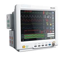
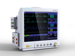
Blood cell analyzer is one of the most commonly used instruments in clinical laboratory. The application of blood cell analyzer not only improves the work efficiency and quality, but also provides more clinical values for the clinical, which is of great significance for the diagnosis and treatment of the disease. There are still some limitations in the process of the extensive application of clinical electrical impedance blood cell analyzer in the process of cell detection, which are often disturbed by some abnormal factors in practical work. This article mainly discusses the interference factors and corrective measures of the specimen in the process of blood cell analysis, so as to ensure the accuracy and reliability of our test results. The common disturbing factors are as follows.
1. common factors of interference
1.1 condensation sample: cold agglutinin disease is autoimmune disease caused by immunoglobulin M (IgM) antibody. It is characterized by the agglutination of red blood cells from patients with certain diseases at lower temperatures. Blood cold agglutination has great influence on the detection of RBC and its parameters. Research shows that cold agglutinin can lead to WBC, MCV false increase, RBC, HCT, PLT pseudo reduction, especially the RBC, HCT decrease particularly significant, but the relationship between the 3 parameters of RBC, HB, HCT is obviously inconsistent, and thus resulting in MCH, MCHC and other calculations. Corrective action: put the specimen in the 37 degree water bath 30min, immediately detect the machine, and the parameters will basically return to normal.
1.2 fat blood samples: fat blood is the most common interference factor. Because the principle of hemoglobin detection is colorimetric method, the factors that cause the increase of serum turbidity, such as hyperlipidemia, abnormal plasma protein, WBC>50*109/L and so on, can lead to the false increase of the serum, which can lead to the significant increase of MCH and MCHC. Treatment: slowly centrifugation, extract plasma and add the same amount of diluent.
1.3 the interference of immunoglobulin: the increase in plasma IgM in Mega globulin and multiple myeloma. High concentration of immunoglobulin interferes with the role of hemolytic agents and causes a false increase in MCHC. Solution: dilute the sample or extract plasma to add the same amount of diluent.
1.4 hemolytic specimens: normal serum free plasma hemoglobin normal reference range: 10-40mg/L, with the hemoglobin in red blood cells, no increase in hemoglobin in red blood cells. However, in pathological conditions, some factors such as DIC, drug, blood transfusion, or blood extraction can induce hemolysis, and the increase of free hemoglobin in plasma can increase the pseudoplasma of MCHC and decrease the RBC. Corrective action: observe whether there are more RBC fragments in the blood smear, or relate to the clinic and re collect specimens.

Meilun The interfering factors of 2.WBC
2.1 nuclear RBC: because the number of RBC in normal cases is about 1000 times of WBC, the total number of leukocytes and the total number of lymphocytes and their proportion are significantly higher [2]. Especially in some diseases, such as hemolytic anemia, a large number of nucleated RBC can be found in peripheral blood, which can not be destroyed by diluent and make WBC count higher. Some five classified blood cell analyzer will suggest the presence of nuclear RBC, and some five classification instruments (MINDRAY BC-6800/6900, etc.) also have an accounting number of nuclear RBC and automatic correction to directly give the exact WBC count result [3]. Corrective actions: manually classify and count the number of nuclear RBC encountered in 100 WBC and calculate according to WBC correction formula.
2.2 large PLT specimens: the blood of some blood system disease patients may have a huge PLT that is the same size as WBC, and these huge PLT are counted as WBC, making the WBC false increase. Corrective action: manual counting of WBC.
2.3 difficult hemolysis or hemolysis samples: severe liver disease due to RBC membrane anti solubility enhancement. Correction: manual counting of WBC; interference with some undisturbed instruments.
2.4 high concentration bilirubin: high concentration of bilirubin affects the count and clustering of WBC. When the free bilirubin is 192.1 u mol/L, the effect on the counting and grouping of WBC is very small, the effect is significant at 256.1 mol/L, and the number of WBC is significantly affected when 384.1 u mol/L, and the instrument can not group [4]. Correction method: use manual method to count WBC.
The interfering factors of 3.PLT
3.1 small cell anemia specimens: small cells in patients with iron deficiency anemia and thalassemia have significant interference with the count of certain instruments PLT. The general impedance analyzer hematology analyzer sets particles of 30-36fL or particles to 60fl (part of optical instrument) to PLT. The impedance hemocyte analyzer can only recognize particle size, and can not distinguish granule properties. When MCV < 70fl, the PLT count of the hematology analyzer is susceptible to interference by small cells and falsely increased. Some high-grade blood cell analyzer can resist small red blood cell interference. The main reason is that these instruments use the optical flow cytology counting principle to detect PLT correction measures: using micromirrors to recount PLT or using optical flow cytometry to detect platelets.
3.2 PLT aggregation: (1) when platelets fail or fail, platelet destruction and aggregation can make platelet count falsely reduce. Measures: re collect blood or manually count PLT. (2) EDTA-K2 anticoagulation, PLT morphology gradually becomes spherical, volume increases, and can cause PLT aggregation. The blood analyzer can not be identified, resulting in a false decrease in PLT. Measures: use EDTA anticoagulant diluent immediately to dilute specimens or microscope to re count PLT, and observe whether there is PLT accumulation in blood smear.
3.3PLT satellite phenomenon: PLT induced by EDTA anticoagulant surrounds neutrophils or lymphocytes / lymphoma.