Wechat QR code

TEL:400-654-1200
TEL:400-654-1200

The basic knowledge of the monitor
1. According to structural classification
The monitoring instruments can be classified into three categories according to their structure: portable monitor, general monitor and telemetry monitor.
(1) portable monitor. The portable machine is easy to carry, simple in structure, stable in performance, can be carried with the battery. It can be powered by battery. It can be used for about 2 hours. It is usually used in the guardianship of non - guardianship and outgoing patients.
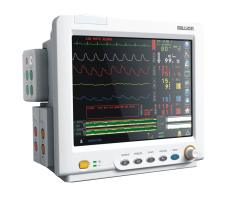
(2) general monitor. General monitors usually refer to bedside monitors, which are more common and widely used in ICU and CHD. It often forms a system with central monitor to monitor.
(3) telemetry monitor. Telemetry is suitable for ambulatory patients and belongs to wireless mode.
2. According to functional classification
Meilun According to the functional classification, there are three kinds of bedside monitors, the central monitor and the ward monitor.
(1) the bedside monitor. It is an instrument connected with the patient at the edge of the sick bed. It can detect the patient's various physiological parameters or some state continuously, and display the alarm or record. It can also work with the central monitor.
(2) central monitor. It can also be called the central monitoring system. It is composed of a main monitor and a number of bedside monitors. It can control the work of each bedside monitor through the main monitor, and monitor the situation of a number of monitored objects at the same time. One of its important tasks is to complete the automatic recording of various abnormal physiological parameters.
(3) away from the hospital monitor. It is generally a small electronic monitor that the patient can carry with him. It can carry out a continuous monitoring of some physiological parameters of the patient in and outside the hospital for a non real time examination.
3. Measuring methods and measuring principles of physiological parameters of guardianship

(I) electrocardiogram
1, the electrocardiogram, also can be called the heart wave.
Electrocardiogram is a curve of the change of cardiac potential recorded from the body surface. It reflects the change of biological potential in the process of cardiac excitation, conduction and recovery.
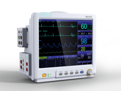
The following is a typical ECG waveform. Each complete ECG waveform contains 6 waveforms: P wave, Q wave, R wave, S wave, T wave and U wave.
The horizontal axis on the recording paper represents time, each 1mm represents 0.04 seconds (the standard paper speed is 25mm/s); the longitudinal axis represents the amplitude of the waveform, and each 1mm represents 0.1mV (when the standard sensitivity is 10mm/mV). Each waveform has its fixed amplitude and time interval. In clinical diagnosis, we can read the electrocardiogram to find out whether it is ill or not.
For example, the normal amplitude of P wave should be 0.2mV, that is, the height of the first wave on print paper should not exceed two grids. By analyzing the ECG waveform, we can find various heart diseases of the heart.
2. The lead.
In order to unify and compare the electrocardiogram, the electrode position of the electrocardiogram and the connection mode of the lead and the amplifier are strictly unified. The way of connecting this electrode group with the amplifier is called the lead or lead of the electrocardiogram.
Now the standard twelve lead is widely used, namely, I, II, III, aVR, aVL, aVF and V1~V6. In general, there is no essential difference between the various leads, but only from different angles to reflect the potential changes of the heart.
The following diagram is the standard connection of the cardiac conductance line.
3. Heart rate
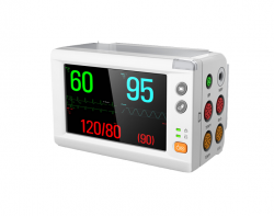
Heart rate refers to the number of pulsations per minute of the heart. There was a significant difference in heart rate between normal adults and quiet time, with an average of 75 times / minute (between 60 and 100 times / min). Heart rate can be different from age, sex and other physiological conditions. The heart rate of the newborn child is very fast, up to 130 times / minute. In adults, the heart rate of women is generally slightly faster than that of men. In the same person, the heart rate slows down during quiet or sleep, and the heart rate increases during exercise or emotional excitement, which can accelerate or slow down the heart rate under the influence of certain drugs or neurophysical factors. People who regularly perform physical labor and physical exercise usually have slower heart rates.
(two) respiratory part
Breathing refers to the patient's respiratory rate, that is, the rate of respiration. Respiratory rate is the number of times the patient breathes in unit time. When quiet breathing, neonatal 60~70 times / points, adult 12~18 times / points. In quiet state, breathing movement was uniform at 16~20 times / minute, with a ratio of 1:4. Male and children are mainly abdominal breathing, while females are mainly chest breathing.
The measurement of respiratory rate in guardianship is impedance.
When the body is breathing, the chest muscle is alternately changed, the chest is alternately deformed, and the impedance of the body tissue alternates. Respiratory impedance (pulmonary impedance) has a certain relationship with lung capacity, and lung impedance increases with the increase of lung capacity. Impedance respiratory measurement is designed according to the change of pulmonary impedance. In monitoring and measuring, respiratory impedance electrodes and electrocardiogram clicks are combined, that is, ECG signals and respiratory impedance are simultaneously detected by ECG electrodes.
The clinical significance of respiration
(1) tachycardia: respiratory rate exceeding 24 times / minute rate of tachycardia. It can be seen in fever, pain, anemia, hyperthyroidism, heart failure, etc. the general body temperature rises by 1 degrees, and the respiration increases by about 4 times / minute.
(2) bradycardia: respiratory rate below 12 times / minute. Slow breathing is seen in excessive anesthetics or sedatives and increased intracranial pressure.